Monday, January 09, 2006

SLT: A New Treatment for Glaucoma Becomes Available

An edited version of this column was published in OCULAR SURGERY NEWS on May 15, 2001

Technology Update

Irving J. Arons
Managing Director
Spectrum Consulting

Introduction

The FDA has recently cleared SLT (selective laser trabeculoplasty) for the treatment of primary open-angle glaucoma (OAG). Coherent Medical (Santa Clara, CA), soon to be combined with ESC Medical Systems(Yokneam, Israel) in a new company to be called Lumenis, will roll out their new Selecta 7000 laser for performing this potentially first-line therapy for primary OAG at the Spring ASCRS meeting in San Diego. The new safer alternative to ALT (argon laser trabeculoplasty), and for non-compliant patients on medical therapy, was cleared by the FDA late in March, 2001. Its key features are its minimal damage to surrounding tissue, long-lasting non-thermal effect in lowering IOP, and repeatability!

Unlike the sometime short-lived thermal effects of ALT in lowering IOP, selective laser trabeculoplasty uses a Q-switched doubled YAG laser to selectively target melanin pigmentation in trabecular meshwork cells, in a process known as selective photothermolysis, thereby delivering no damage to non-targeted adjacent tissue. Because SLT is non-thermal, it is repeatable, a distinct advantage over ALT. For this reason alone, not withstanding its clinically-proven long-term effect in lowering IOP (up to 80% success in obtaining a mean 23% reduction in IOP lasting for at least 24 months in international trials for patients on maximally tolerated medical therapy, and still uncontrolled), it could have a significant impact on glaucoma therapy, possibly becoming the first approach to treatment.

Underlying Principles and History

Dr. Mark Latina, the inventor or SLT, first became interested in selectively targeting trabecular meshwork cells while a fellow at the Boston-based Mass Eye & Ear Hospital in 1985, and later, working under an NIH grant, transferred his work to the Wellman Laboratories of Photomedicine at Mass General Hospital in 1987. Work was underway at the Wellman at that time on discovering the benefits of selective photothermolysis, discovered by Drs. Rox Anderson and John Parrish in the early 1980s. Selective photothermolysis, now used extensively in the laser treatment of several skin diseases and in laser-based hair removal, is based on three principles. First, absorption of intracellular targets must be greater than that of surrounding tissues (such as melanin as a chromophore in the trabecular meshwork). Second, a short pulse of laser energy is required to generate and confine heat to the pigmented targets, with the wavelength matching the absorption wavelength of the target. (Again, melanin proved a worthy target.) Finally, the pulse duration must be less than or equal to the thermal relaxation time of the target. When all of these conditions are met, target specificity becomes independent of focus.

Based on some published observations of Jorge Alvarado, MD, pertaining to the decreased cellularity in the trabecular meshwork, Dr. Latina and his colleagues at Wellman attempted to separate the coagulative effects from the purely biological effects, as well as the physiological effects on the TM of laser trabeculoplasty, and to see if the cells could be selectively targeted. At that time (in 1985/1986), two types of lasers were in use in ophthalmology, continuous wave argon, for thermal coagulation, and the Q-switched, pulsed Nd:YAG, just coming into use for photodisruption (optical breakdown) of secondary capsules, and for iridotomy. The conventional argon laser trabeculoplasty (ALT) used a pulse generation of 100 ms to transfer heat to the focal spot in the trabecular meshwork, causing a coagulative burn and creating a scar, which helped to improve fluid outflow through the shrunken tissue surrounding the scar. With the work underway at Wellman on confining thermal damage to selected pigmented targets, Latina and his colleagues thought that if the appropriate laser parameters were chosen, a specific type of laser tissue interaction could be achieved. And thus, after experimentation with various short-pulsed lasers, and using the melanin pigment in the TM cells as a target in cell viability cytotoxicity assays, it was discovered that a single pulse of a Q-switched 532 nm doubled YAG laser could effect only the pigmented TM cells aimed at, while not effecting pigmented cells or other structures outside of the irradiation zone, and most importantly, there was no gross disruption of the TM cells being targeted.

With the argon laser, there was no region where selective killing could be achieved, even with short pulse durations. However, with the 532 nm Q-switched laser, selective targeting was achieved using threshold fluences of only 50 mJ/cm2. This was a much lower fluence that the 107 mJ/cm2 then in use with the conventional Nd:YAG 1064 nm photodisruptive laser. A 400 um spot size is used with the 532 nm laser for SLT, which is quite large compared to the typical 10 um spot used with the Nd:YAG (or 50 um spot used with the argon laser in ALT). Thus pumping 1 mJ of energy into the 10 um spot results in extremely high irradiances, while the same 1 mJ pumped into the 400 um spot of the SLT laser results in very low energy per unit area. This discovery was patented by Dr. Latina in U.S. Patent 5,549,596.

Beginning about 1993, as part of a support program for work underway at the Wellman Laboratories, Coherent Medical became aware of Dr. Latina's work and began supporting it directly, including building a clinical laser for him. The company modified one of its ophthalmic Nd:YAG lasers, adding the doubling crystals to bring the wavelength down to 532 nm, and added the optics necessary to create the 400 um spot. This ultimately became the Selecta 7000, used in both the U.S. and international clinical trials, and now approved by the FDA for treating primarily open angle glaucoma with SLT. Coherent obtained an exclusive license to the SLT patent from Mass General Hospital upon its issue in 1996.

The major advantages of SLT over ALT are: 1) selectivity, only the melanin pigment in TM cells is targeted; 2) no thermal damage or gross disruption of the TM cell architecture (and thus excellent safety); and 3) the potential for repeatability of the process. Unlike ALT that creates scars in the trabecular meshwork, SLT does not do damage to other than the targeted cells, and thus the treatment can be repeated if further reduction in IOP is needed. With the FDA approval of SLT in late March 2001, HCFA has approved reimbursement equivalent to ALT, or on average of about $385 per procedure. The Selecta 7000, which has the CE mark and has been available for sale in the European Union since 1998, and is approved by the Ministry of Health for sale in Japan, will be commercially launched in the U.S. at the Spring ASCRS meeting, priced at $55,000.

Technique and Clinical Trial Data

The procedure for use is similar to a conventional ALT. Preoperatively, careful gonioscopy is done to visualize the trabecular meshwork and plan the treatment area. A drop of each of Iopidine or Alphagan and topical anesthesia are applied, and a Goldman three-mirror goniolens is placed on the eye with methylcellulose. The aiming beam is focused onto the pigmented trabecular meshwork and the treatment is done by placing 50±5 contiguous but not overlapping 400 um single laser spots along an 180° treatment arc. Unlike argon laser trabeculoplasty, there are no visible coagulation burns to the trabecular meshwork. If bubble formation occurs, it means the pulse energy is too high. Bubble formation is monitored with each pulse. After laser treatment, prednisolone acetate 1% is administered and continued in the treated eye four times daily for four to seven days.

Selective trabeculoplasty is not associated with coagulation damage, but significantly lowers IOP. This indicates that coagulation of the TM structure is not important to the mechanism of IOP-lowering. It is believed that SLT works on the cellular level, either through migration and phagocytosis of TM debris by the macrophages, or by stimulation of formation of healthy trabecular tissue which may enhance the outflow properties of the TM.

Beginning in 1998, a prospective, U.S. multicenter pilot study was conducted at three sites for evaluating the IOP-lowering effect of SLT. Fifty-three eyes of 53 patients whose IOP was not controlled with maximum medical therapy, the OAG group; and a second group of 67 patients with uncontrolled OAG who had previously failed argon laser trabeculoplasty, the post-ALT group, were included. Most patients had primary OAG, a few had pseudoexfoliative glaucoma, and a further few had other types of the disease. All anti-glaucoma medications were maintained during the treatment and follow-up periods. Of the 120 patients treated with SLT, 101 (84%) were evaluated in terms of efficacy analysis for the FDA. Fifteen patients discontinued the study early for various reasons. The remaining patients were followed for 26 weeks postoperativelyy.

The results showed that 72% of both SLT treatment groups of patients were responders. The average preoperative baseline IOP was 25.4 mm Hg, and showed a mean IOP reduction of 5.9 mm Hg (23%) at 26 weeks. In the OAG group, the mean baseline was 25.1 mm Hg. These patients showed an IOP reduction of 6.0 mm Hg, or a 23.9% drop. The post-ALT group was slightly closer in both the responder rate as well as IOP reduction overall, with 71.4% responding with an IOP reduction of 5.8 mm Hg. The untreated eyes had an average preoperative IOP of 21.2 mm Hg and had a reduction of 2.8 mm Hg (13.2%). SLT was equally effective in IOP reduction as ALT in both the OAG and post-ALT groups. There was no persistent IOP elevation following the SLT treatment in the post-ALT group, which is prevalent following ALT treatment.

Adverse effects consisted of peripheral anterior synechiae in 8 patients (6.7%) of treated eyes, one in the OAG group and seven in the post-ALT group. (Seven patients also were reported to have peripheral anterior synechiae in the untreated eyes.) Anterior chamber inflammation with observable cells and flare was noted in approximately 80% of eyes in the early postoperative period. The inflammation was treated with topical steroids and dissipated within 24-72 hours. Fifteen percent of patients complained of minimal pain, discomfort, and redness during treatment or at follow-up.

A prospective, randomized Canadian clinical study by Dr. Karim Damji compared SLT with ALT in 36 eyes. Baseline IOP was 22.8 mm Hg and the SLT group had 22.5 mm Hg. For the ALT group, IOP reductions were 4.8 mm Hg at 6 months, while the SLT group had an average reduction of 5.0 mm Hg.

The largest group of patients treated to date were in Germany, where Drs. Weimer, and Kaulen investigated 460 eyes with 2 years of follow-up. The SLT-treated group had a mean decrease in IOP of 23%, with a two-year success rate of 80%. The complication rate was roughly 4.5%, much lower than the complication rate for ALT, which can reach 34%. The most common complications were elevation of IOP (2.4%), and significant inflammatory reaction in the anterior chamber without an IOP spike (1.5%). All complications were easily treated.

Conclusion

SLT is as effective as ALT in the treatment of primary OAG patients. SLT appears to be repeatable, because of the lack of coagulation damage and the demonstrated efficacy in patients with previously failed ALT treatments. With this lack of tissue damage, SLT should be considered, and has the potential to evolve as an ideal primary treatment option in open angle glaucoma patients who cannot tolerate or are non-compliant with medications, without interfering with the success of future surgery.

Avastin: A New Hope for Treating AMD

This report was prepared for publication in my Journal on December 31, 2005.

Irving J. Arons
Spectrum Consulting

Over the years, beginning in December 1994 (1-3), I have been reporting on potential laser-based treatments for age-related macular degeneration (AMD); primarily because my specialty was laser technologies and not drugs/pharmaceuticals. However, this past summer, my eyes were opened, as I read a review by Lynne Peterson in Trends-in-Medicine, reporting on what she learned at the July 2005 American Society of Retina Specialists meeting (4), held in Montreal.

First Reports about Avastin

Lynne reported about two drugs from Genentech — Lucentis and — for the first time — its sister drug, Avistan. Both are anti-VEGF agents and have the ability, not only to stop wet AMD cold in its tracks, but to actually improve vision for those afflicted with the disease.

As she wrote, “Word spread like a tsunami through the American Society of Retina Specialists (ASRS) meeting about the newly discovered benefits in wet age-related macular degeneration (AMD) from the off-label – and very inexpensive – use of a chemotherapy agent for colorectal cancer. At the beginning of the meeting, only a handful of doctors knew about intravitreal injections of Genentech’s Avastin (bevacizumab), but by the end of the meeting, most doctors questioned said they plan to go home and try it.”

“In fact, Avastin stole the show from Genentech’s Lucentis (ranibizumab), a fragment of the Avastin molecule that is being developed specifically as an intravitreal injection for AMD. The data presented on Lucentis was outstanding, but it did as much to convince doctors of the value of Avastin as to build anticipation for Lucentis. There have been no studies of Avastin, just case reports and personal experiences, but that was enough to make doctors want to use it – particularly in patients who have failed photodynamic therapy and/or Eyetech’s Macugen (pegaptanib).”

First Reports from the AAO

This piqued my interest, which was soon aroused again as the first reports from the October 2005 American Academy of Ophthalmology (AAO) meeting emerged, especially from the Retina Subspecialty Days presentations. The first report I read was Dave Harmon’s AAO report in his November Ophthalmic Market Perspectives (5), in which his #1 key area of directional shift in the thinking of ophthalmologists was The Avastin Embrace. As he put it, “After years and countless millions spent in development of a new generation of AMD drugs, many of which are still in final stages of FDA trials, the retinal community is most excited about the results from treatment with a low cost drug now designated as a treatment for colorectal cancer. If the Avastin momentum continues, it could reduce the cost of AMD treatment to a small fraction of current amounts and wrench the wind from dozens of new drug development programs.”

Later, in his report on the Retina Subspecialty Day, he wrote: “Record attendance at the Retina Subspecialty Day Meeting underscored excitement and interest in new treatments for retinal disease. Updated clinical data on new drugs and therapies for treatment of AMD garnered the most interest at the two-day meeting. The agenda included the latest clinical data for new drug therapies including Macugen (EyeTech Pfizer), Lucentis (Genentech), and Retaane (Alcon). Also featured were updates in clinical experience with photodynamic therapy (PDT) and several studies of combination PDT and drug therapy.

Lucentis continues to be the most exciting new drug in the development channel. Jeffery Heir, MD, reported that one-year patient data from the Lucentis Marina clinical trial showed that approximately 95 percent of patients treated with the drug lost fewer than 15 letters of visual acuity compared with 62 percent in the control group. On average the patients treated with Lucentis had a significant improvement in visual acuity relative to baseline, whereas the control group experienced a substantial decrease in visual acuity from baseline.

However, another Genentech drug generated the most excitement. Avastin, of which Lucentis is a fragment, has been used in the treatment of colorectal cancer since 2004, and works by blocking the formation of new blood vessels leading to metastasis. Phillip Rosenfeld, MD, PhD, of the Bascom Palmer Eye Institute reported on treatment results using systemic Avastin in salvage patients (patients who had not responded to other drugs). In the spring of 2004, researchers initiated a Phase I/II trial using Avastin which now includes 18 patients followed for 24 months. The results appear to be similar to those of Genentech’s Lucentis, with AMD patients experiencing improvements in vision...

George Williams, MD, cautioned attendees on the impact of high-priced AMD treatments for US patients. Williams reported that with the currently approved medications, the annual cost of AMD therapy in the US could reach $1 billion. While Medicare will cover the majority of these costs, current policies designed to reduce expenses will lead to a 40 percent decrease in Medicare reimbursement for treatment of AMD by 2013. Williams said that this situation places a significant burden on retinal specialists to select treatments that are in the best interest of the patient and society as a whole. “

This was quickly followed by Michael Lachman (of Lachman Consulting LLC) in his EyeQ Report No 2 (6) of the AAO meeting, in which he reported that (the treatment of) AMD was in transition.

As he put it, “Treatment regimens for wet age-related macular degeneration (AMD) are about one year into what will be a period of transition lasting three years or more. With Eyetech and Pfizer’s Macugen about ten months into its US launch, sales have risen steadily and have had a meaningful impact on domestic Visudyne sales. Visudyne is holding up well internationally, where Macugen is generally not yet available. Off-label use of Genentech’s Avastin is also having an impact on both Macugen and Visudyne sales in the US. Meanwhile, Genentech just released impressive clinical results for Lucentis from the pivotal ANCHOR trial, to go along with the previously released impressive results from the pivotal MARINA trial. Genentech plans to file its Biologics License Application (BLA) next month and request priority review. Once Lucentis is approved, as early as H2-06, the impact on both Visudyne and Macugen will be immediate and significant.

The next report that caught my eye was the supplement that accompanied the December 2005 issue of Review of Ophthalmology, entitled AMD 2005: Evolving treatment Options (7), especially the update on Bevacizumab (Avastin) by Philip Rosenfeld, MD, Associate Professor of Ophthalmology at Bascom Palmer Eye Institute.

He wrote about anti-VEGF drugs and Avastin in particular. (He is the foremost investigator into this drug and its applications in treating wet AMD.) But the telling point he made is about the cost differential between Avastin and the other two anti-VEGF drugs, Macugen and Lucentis. It appears that Avastin, which is already FDA approved for use in treating colon cancer, costs about $5.50 per dose, compared to $1000 - $3300 per dose for Macugen (or Lucentis, when it is approved sometime later this year).

On December 30, 2005, Genentech filed for a Biologics License Application (BLA) of Lucentis (8), asking for “priority review” approval, which could come in as little as six months.

Clinical Results

Finally, a report came across my desk from the recent Hawaiian Eye and Retina 2006 meetings held in January in Hawaii. Michael Lachman issued another EyeQ Report (9) stating, More Good News for AMD Patients (and Presbyopes). Two parts of Michael’s report were his highlights: “Highlights of the Retina 2006 program: (1) presentation of additional outrageous Phase III data for Genentech’s Lucentis for wet AMD, (2) growing off-label use of intravitreal Avastin for a number of retinal indications, and (3) diminishing interest in Macugen, Visudyne/PDT, and intravitreal triamcinolone”... and his report on the one-year results of the Genentech pivotal ANCHOR study, comparing Lucentis at two dosage levels to Visudyne/PDT as the control.

The results for Lucentis were outstanding, with Lucentis actually showing significant improvement in vision of 36% to 40%, compared to 6% for Visudyne.

Of the primary endpoint, 94% and 96% of Lucentis patients maintained vision (i.e., a loss of less than 3 lines of visual acuity (VA)) compared to 64% with Visudyne.

Of the secondary endpoints, the aforementioned 3+ lines of vision was achieved for 36% and 40% with Lucentis and only 6% with Visudyne. Changes in VA of 20/40 pre-op to post-op went from 1.4% to 31.4% for 0.3 mg doses and 4.3% to 38.6% for the 0.5 mg dose. Meanwhile, the Visudyne controls only improved from 0% to 2.8%!

There were other significant changes, including severe loss of vision of 0% with Lucentis compared to 13.3% with Visudyne. (For the rest of the results, please see the table on page 2 of the EyeQ Report, and shown below.)


Commenting on the off-label use of Avastin, Lachman noted that since the first announcement by Rosenfeld at the ASRS in Montreal in July, over 5000 invitravitreal injections have been administered to patients in the U.S.

Lachman also commented on the anecdotal uses of both drugs by some physicians who believe that Lucentis might be slightly more effective than Avastin.

The fly in the ointment, however, might depend on reimbursement. As Lachman further noted, “CMS is cracking down on physician reimbursement for intravitreal injections of Avastin. The cost of the drug itself is not reimbursed, although in quantities used for intravitreal injection, the cost is relatively low at $15-75 per dose. Based on the investigational nature of intravitreal Avastin and the lack of supporting clinical data, all 17 Medicare carriers covering all 50 states have issued verbal denials of coverage, and seven carriers covering 12 states have issued written denials. Retina specialists are hoping that the reimbursement situation will improve in the coming months, supported by the near-term publication of a number of peer-reviewed articles, in RETINA and other journals, covering physician-led studies of intravitreal Avastin.”

The Bottom Line

Advances in the treatment of wet AMD are definitely on the horizon. Improvements over current treatments are possible today with the off-label use of Avastin — although reimbursement is definitely a problem, while Lucentis will probably be approved within six months and, will be reimbursable (although at a much greater cost to the Medicare system).

As new events unfold in this story, I will attempt to update the status of both Lucentis and Avastin.

References:

1. “Photodynamic Therapy for Macular Degeneration”, I.J. Arons, Technology Update, Ocular Surgery News, December 15, 1994.

2. “Laser Treatments for AMD Show Promise”, I.J. Arons, Technology Update, Ocular Surgery News, January 15, 2000.

3. “Visudyne Looks Promising for Treating Wet AMD”, I.J. Arons, Retina/Vitreous, Ocular Surgery News, January 15, 2001.

4. “American Society of Retina Specialists, Montreal Canada, July 16-20, 2005", Lynne Peterson, Trends-in-Medicine, July 2005.

5. “New Directions Set at 2005 AAO Meeting”, David Harmon and William Freeman, Ophthalmic Market Perspectives, Market Scope, November 11, 2005.

6. “Ophthalmology Q3-05: IOLs Strong, Refractive Mixed, and AMD in Transition”, Michael Lachman, EyeQ Report No. 2, Lachman Consulting LLC, November 18, 2005.

7. “AMD 2005: Evolving Treatment Options”, Supplement to Review of Ophthalmology, December 2005.

8. “Genentech Announces Filing of BLA for Lucentis”, PRNewswire, December 30, 2005.

9. “Hawaiian Eye and Retina 2006: More Good News for AMD Patients and Presbyopes”, Michael Lachman, EyeQ Report No. 3, Lachman Consulting LLC, January 23, 2005.

Note: Some of the references above, notably the report from the ASRS meeting in Montreal (4) and Dave Harmon's report from the AAO (5) are subscription only. Anyone who would like to see the specific references are urged to email me and I will forward you a saved copy.

Author’s Note on Avastin

Since the original posting on January 31st, I have added three updates on this important drug for treating age-related macular degeneration. In addition to the posting you are reading, here is a listing (with links) to the others:

Avastin Update: Medicare not Likely to Cover its Use

Avistin Update II: AAO supports Medicare Coverage for Off-label Avistan Use

ARVO 2006: A Further Update on Both Avastin and Lucentis for Treating AMD

Laser Treatments for AMD: Visudyne Looks Promising for Preventing Blindness

An edited version of this column was published in Ocular Surgery News on January 15, 2001.

Irving J. Arons
Managing Director
Spectrum Consulting

As I reported following the 1999 AAO meeting ("Laser treatments for AMD show promise", OSN, Jan. 15, 2000), Visudyne PDT therapy appeared promising for those afflicted with predominately classic, wet age-related macular degeneration (AMD), along with several other PDT treatments in the pipeline. One of the questions posed by several industry analysts during that meeting was how, when these new techniques came to market, would HCFA handle the reimbursement issues without bankrupting Medicare?

Well, the inevitable has happened, Visudyne therapy (verteporfin for injection, marketed for QLT, Inc. by CIBA Vision) for predominantly "classic" wet AMD was approved for marketing by the FDA in April 2000. Since then, literally thousands of people with AMD have flooded retinal surgeon's offices seeking treatment. The approval was based on the 12-month data from two, 24-month randomized, double-masked, placebo-controlled Phase III clinical trials known as the TAP (Treatment of AMD with Photodynamic therapy) Investigation. The results of the TAP studies were published in the October 1999 issue of Archives of Ophthalmology. Basically, TAP 12 month findings showed that in 243 patients with predominantly classic wet choroidal neovascularization (CNV), vision remained stable or improved in 67% of patients treated with Visudyne therapy compared to 39% of patients in the placebo arm of the study. Additional data released this past summer showed that the beneficial effect and the favorable safety profile of Visudyne therapy, observed at the 12-month time point, was maintained out to two years, with fewer treatments required in the second year (see the clinical results update below).

Market Size and Reimbursement Issues

It is now estimated that some 65,000 U.S. treatments, both primary and retreatments (90,000 worldwide), were done in 2000, producing between $90 million to $100 million in drug revenues for QLT/CIBA Vision. And, if analysts projections continue to hold up, the drug could reach over $200 million in sales in 2001 -- and as high as $500 to $700 million in revenues by 2003.

But the picture isn't all rosy. In November, HCFA issued its revised National Policy for Reimbursement, calling Visudyne therapy a "medically reasonable and necessary treatment". The agency also expanded somewhat, the number of patients who can receive treatment -- those presenting with at least 50% classic symptoms from a fluorescein angiogram; and how often retreatments can occur -- up to 6 treatments over 24 months, although recent field interviews/surveys are showing that fewer retreatments are occurring in practice and/or are over an extended time frame. (The TAP inclusion criteria included having visual acuities between 20/40 to 20/200, and lesions greater in size than 0.5 mm. Also, on average, 5.6 treatments were reported over the two year period.)

However, the agency cut the expected physician reimbursement for the treatment, including infusion, use of the laser, and staff overheads, to $341 from the $500 to $700 reimbursement figure expected (and previously paid for laser photocoagulation). When added to the drug reimbursement ($1458), Visudyne PDT therapy total reimbursement will be $1799 in 2001. Taken against the cost to administer the treatment, according to a Dain Rauscher Wessels analysis, a high volume practice could expect a profit per procedure of about $500, while a low volume practice might only clear $135 -- and that does not include the physician's fee! This total is down from the $900 profit that a high volume practices could have expected under the older laser photocoagulation rates, and about $550 for lower volume practices.

With between 1200 to 1300 practicing retinal specialists in the U.S., we estimate that there are currently about 600 PDT activation lasers (from Zeiss Humphrey and Coherent Medical) in use in the United States (with an additional 600 in use in the rest of the world).

When queried, in a poll of 67 retinal practices undertaken by Leerink Swann & Company in November 2000, 82% of respondents stated that the new level of reimbursement from HCFA was below expectations, and inadequate. At an AMD press conference held during this years Academy meeting, prior to the HCFA announcement, Dr. Mark Blumenkranz noted that Visudyne treatment may end up being offered only at larger medical centers or academic centers if the reimbursement issues are not satisfactorily resolved.

AMD is the leading cause of severe vision loss in the elderly in the U.S. The National Institute of Health (NIH) estimates that nearly 1.7 million elderly Americans, 5% of the total population over 65 years of age, have some degree of vision loss due to AMD.

I have attempted to calculate what the cost to the Medicare system for PDT might be for the year 2000 and beyond. The cost for 2000 depends on how many people actually underwent the treatment, which, in turn, depends on how many of the 200,000 to 350,000 of the current AMD pool met the acceptance criteria. The wet form of AMD accounts for approximately 10%-15% of the total AMD population, or between 1.3-1.5 million people in the United States, plus another 200,000 new cases diagnosed each year (and an additional 400,000 in the rest of the world). The predominately classic form represents approximately 10% to 15% of the total wet form population, or approximately 130,000 to 250,000 people that could now be treated, with an additional 20,000 to 30,000 being diagnosed and entering the potential treatment pool each year, not taking into account additional patients with diseases such as pathological myopia, which should be added to the Visudyne label sometime in mid-2001.

The cost to the Medicare system for Visudyne PDT in 2000 could conceivably have ranged from $160 million to $280 million, assuming that one-third of the current pool had elected for treatment, one-half of those were eligible (many in the pool may have already lost too much vision to be treatable), and the cost to the system is $1799 per procedure ($1458 for the drug plus $341 for treatment) with an average of three treatments needed over the course of the first year. (The actual cost for 2000, based on 65,000 doses, was closer to $115 million.) In subsequent years, with another third of those eligible entering treatment, along with additional retreatments for some of those already started on their initial course of treatment, Medicare costs could average over $250 million a year. And when other eye diseases and treatment modalities are approved, the cost can only go upward. Another factor to consider is the subjective nature of the diagnosis. Those patients with marginal classic symptoms may be included in the treatment class, as this might be their only hope for retaining vision.

However, on a more positive note, Dr. Sanjay Sharma of Queens University in Kingston, Ontario, presented a paper during the Academy meeting on a decision making model for measuring the impact of Visudyne therapy on the quality of life of those with AMD. He concluded that PDT is a very effective treatment for AMD, and that a patient who is still able to drive, could expect a 10.7% improvement in their quality of life if they received the treatment, while patients who were legally blind could expect a 7.8% improvement. This compares to those who have lost significant vision, and who are assessed as having a 40% reduction in their quality of life. With the looming crisis in vision loss as baby boomers advance into their mid- and senior years over the next decade, more than 500,000 people a year will be diagnosed with AMD, and without treatment, will become more dependent on others (and possibly the welfare system). As his model suggests, "For one patient with macular degeneration to obtain one quality of life-adjusted year, photodynamic therapy will cost a managed care organization $86,721. If the treatment proves effective over the entire duration of a patient's life, this cost will fall dramatically." And what is left unsaid, is what the cost to the system would be for legally blind people, not able to avail themselves of the treatment!

Clinical Update - Wet AMD

PDT Trials

During the AAO Vitreo-retinal Update pre-meeting, Retina 2000, Dr. Susan Bressler provided the 24 month data for the TAP study. The data re-iterated the significant difference in those eyes treated with PDT versus the placebo control, with 53% of the Visudyne patients losing less than 15 letters (three lines) of visual acuity, compared to 38% for the placebo group. The sub-group results for predominately classic patients showed even better results, but were not as good for those with minimally classic, or non-classic, disease. For those with predominately classic lesions, 59% lost less than 15 lines, versus 31% of the placebo group; in the minimally classic group, 48% fared better than 44% getting the placebo treatment; and 56% versus 30% for the non-classic disease group. Dr. Bressler concluded that the results seen after 12 months were sustained for the 24 months, with even more compelling evidence to use Visudyne therapy for patients with classic wet AMD. The additional benefits of the 24 month TAP trial were limited, but included; slower lesion growth, reduced leakage, and stable contrast sensitivity. The average number of treatments of the group during the second year was 2.2 out of a possible maximum of 4, resulting in the average number of treatments over the two-year period as 5.6 out of a maximum of 8.

Dr. Joan Miller reported on the 12 month results of the Verteporfin in Photodynamic Therapy (VIP) study for pathological myopia, taking place at 28 clinical centers worldwide. In this case, 72% of those treated achieved less than 8 letter loss (less than 1.5 lines) compared to 44% in the placebo group. Dr. Miller concluded that Visudyne treatment resulted in a significantly increased incidence of stability to improved visual acuity, with no evidence of ocular or systemic tissue risk. Compared to the TAP study, the VIP trial was focused on two groups of patients; those with pathologic myopia and those with an earlier stage AMD (i.e. either those with occult lesions or those with classic lesions but better than 20/40 vision). While the VIP trial was intended to expand the population for Visudyne eligible patients, it was less definitive than TAP. There were statistically beneficial benefits experienced among the pathological myopia group, however there was no statistically significant differences/advantages for those with earlier stage AMD treated with Visudyne compared with placebo.

At the Academy's AMD press conference, Dr. Blumenkranz, coordinator for the Pharmacyclics/Alcon Labs Optrin (LuTex) trials, said that the Phase I/II clinical trials had been completed and were being evaluated prior to beginning recruiting patients for Phase III trials. (It was also learned that a Zeiss diode laser is being used for activation, and not the Diomed system as reported in my January 15th article.)

Dr. Edgar Thomas, head of the Miravant/Pharmacia & Upjohn Photopoint (purlytin) trial, said that it was completing Phase III trials, with nothing new to report.

Laser Treatments

Dr. Elias Reichel reported on the results to date in the ongoing multi-center clinical trial to treat occult wet AMD, the most prevalent form of the disease, with low intensity laser energy, in the Transpupillary Thermotherapy (TTT) for CNV trial. Rather than photodynamic therapy, this trial uses Iridex's Iris Medical's SLx 810 nm diode laser in its unique longpulse mode to treat the lesions without the need for a photoactivatable drug. He reported that after one year, 80% of patients with occult wet AMD treated with the laser experience a halt in new vessel growth and that 70% had stable or improved vision, without any signs of damage to the photoreceptor cells of the retina. The TTT treatment appears to stop the evolution of the exudative process, and may avoid the development of or progression to the classic form. (Traditionally, half of all cases of occult AMD move into the classic form, with profound visual loss.)

Anecdotal evidence indicates that the TTT treatment is doing quite well in the field, but the reimbursement picture remains cloudy. According to Iridex, reimbursements for this treatment, which are left up to the discretion of local Medicare carriers, range from a low of $127, to a high of $700. Until this discrepancy and confusion is cleared up, and published peer-reviewed studies on its success begin to appear, TTT treatment will be slow to be accepted by most retinal surgeons.

Clinical Update - Dry AMD

At the Iridex booth, a number of speakers provided updates on the ongoing work with both therapeutic and prophylactic treatments for the dry form of AMD. Based on a pilot study in which their 810 nm diode laser was used in a grid-pattern therapy of non-exudative soft drusen, with resorption of drusen seen in 68% of treated eyes and visual acuity improvement in 24% of a subset of treated eyes after a single treatment, the company continues to sponsor additional work in both therapeutic and prophylactic trials. In the therapeutic study, the four-year followup of the original pilot study showed continued improvement in 78% of treated eyes, defined as a reduction of equal to or greater than 50% of drusen from baseline, versus only 8% of eyes not treated, but observed. This resulted in vision improvement by two or more lines of visual acuity in 14% of treated eyes, which suggests a therapeutic benefit for patients with dry AMD who have lost two or more lines of VA due to the presence of central soft drusen.

In the ongoing prophylactic study, Prophylactic Treatment of AMD (PTAMD), a total of 35 neovascular events have occurred in the four years in the 22% of non-responsive to treatment eyes, with half occurring in the observed eyes and half in the treated eyes, indicating no treatment harm nor benefit. However, in the 78% of eyes that responded to treatment with a significant reduction in drusen, there was only one eye that developed CNV. The study authors suggest that a prophylaxis treatment that effectively promotes drusen resorption may be effective in reducing or delaying progression to CNV.

It is beginning to appear that the once dreaded age-related macular degeneration disease, and the visual loss accompanying it in its worst case, will become treatable for the vast majority of those who contract it over the next decade.

Friday, January 06, 2006

Laser Treaments for AMD Show Promise

An edited version of this column, along with an accompanying table listing the market participants, was published in the January 15, 2000 issue of Ocular Surgery News.

Treatments for AMD, Even Prevention, are on the Horizon

Irving J. Arons
Managing Director
Spectrum Consulting

This year, as reported at the 1999 AAO Meeting in Orlando, for the first time it appears that one or more methods for stopping the progress of age-related macular degeneration (AMD) and, perhaps, even to improve vision for those who suffer from the disease, is on the horizon. And in an even bigger leap of faith, additional clinical work is being undertaken to determine if this terrible disease can be stopped before it progresses to the final stages that causes legal blindness.

What is AMD?

AMD is a progressive degenerative condition that damages the macula, the small central part of the retina in the back of the eye which provides central vision, and currently affects approximately 15 million people in the U.S., and at least double that in the rest of the world. Loss of central vision robs a person of the ability to perform tasks which require fine focus, such as reading, watching TV, or seeing faces. AMD can occur in two forms, the "dry" (non-exudative) form and the "wet" (exudative) form, and both can coexist in the same patient. The dry form, accounting for between 85% to 90% of AMD, is typically characterized by a build-up of drusen, white to yellow deposits underneath the retina in the macula that initially results in minimal visual symptoms. With time, vision may gradually deteriorate, depending upon drusen type, size, quantity, and location. More importantly, the presence of drusen constitutes a risk factor to develop severe vision loss due to the development of geographic atrophy or the development of the exudative (wet) form of AMD, accounting for approximately 10% to 15% of all AMD, but 90% of severe vision loss (legal blindness) in those over the age of 50.

The wet form is typically characterized by a proliferation of abnormal blood vessels (i.e., choroidal neovascularization [CNV]) underneath the macula and results in a more rapid and profound loss of central vision. About 1.5 million to 2.5 million American people (approximately 10% to 15% of all AMD cases) currently have the wet form, which accounts for the vast majority of AMD patients who are legally blind. AMD is the leading cause of blindness in the U.S. for all age groups. For persons 65 to 74 years of age, the prevalence approaches 30%. In the U.S., the current annual incidence of AMD is approximately 2 million people, 200,000 to 300,000 of whom have the wet form. It is expected that both the incidence and prevalence of AMD will increase substantially with the aging of the population.

Treatments for Wet AMD: Laser Photocoagulation

Currently, there is no clinically proven interventional therapy for dry AMD while a minority (about 15%) of patients with the wet form of AMD can be treated using laser photocoagulation, which works to close and stop progression of the bleeding CNV vessels. Photocoagulation usually causes the immediate further loss of visual acuity due to the thermal damage done to the overlying retina, and can only be applied to those patients, with "classic", well-demarcated CNV lesions. In spite of these deficiencies, laser photocoagulation is widely employed, principally because it is the only treatment option available for sufferers of wet AMD. Approximately 42,000 laser photocoagulations were performed last year, used in less than 2% of eyes with AMD. Thus there is a need for a treatment modality suitable for a large segment of AMD patients that selectively closes the choroidal neovessels with out damaging overlying neurosensory retina and without causing further loss of vision. The photodynamic therapy (PDT) approach appears to be one such treatment.

Photodynamic Therapy for Wet AMD

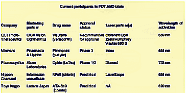
Most of the AMD news to date, has been on the ongoing studies of photodynamic therapy to stop the progress of classic wet AMD, those lesions that are well defined, leak heavily, and cause the most rapid deterioration of vision. Several studies are underway, with the QLT PhotoTherapuetics' team (Ciba Vision for marketing and Coherent and Carl Zeiss with the activating lasers) in the lead. QLT's PMA for Visudyne therapy recently won recommendation for approval from the FDA's Ophthalmic Drug Advisory Panel. Following behind is the Miravant team (Pharmacia & Upjohn as the marketer, and IRIDEX's Iris Medical as the laser supplier), and then Pharmacyclics (with marketing partner Alcon Laboratories, and probably Diomed as the laser supplier). FDA final approval of QLT's Visudyne injection therapy might be in hand before this article reaches print, most likely in the first quarter of 2000. Miravant's Photopoint therapy has completed enrollment of patients in its Phase III clinical trial, with a PMA expected to be filed sometime in 2000 or 2001. Pharmacyclics' Optrin therapy is even further behind, still running Phase I/II clinical trials. (Others in preclinical trials include Nippon Chemical and Toyo Kogyo, in association with Lederle Japan.) (See the table below for a listing of the players and the drugs/lasers involved.)


In the PDT approach, a photosensitizer is administered intravenously, which selectively accumulates in the neovascularized tissue. A dose of carefully calculated laser light is administered directly to the lesions, which activates the photosensitizer causing death to the cells and destroying the vessels. It is important to note that the effect of PDT treatment is photochemical and there is no accompanying thermal reactions. Although the results are a stoppage of blood leakage from the disruption of the vessels, according to the results seen in the TAP study (Treatment of Age-Related Macular Degeneration with Photodynamic Therapy), as reported at the Vitreoretinal Update '99 meeting held just prior to the AAO, the effect appears to be temporary, and as many as four treatments per year may be needed. However, there are indications that the recurrance of bleeding should lessen with time. The problem is the cost of the treatment. Visudyne is expected to be priced at about $1200 per dose, and with the surgeon and facility fee added in, a single treatment could cost as much as $2000, or $8000 per year. But, the results of the TAP study indicate that visual acuity, contrast sensitivity, and angiographic outcomes are better in verteporfin-treated eyes, than those receiving a placebo. Verteporfin therapy can safely reduce the risk of vision loss in eyes with predominantly classic CNV lesions. However, vertoporfin therapy does not appear effective for occult CNV, typically poorly defined and diffuse lesions with less leakage, the most common form, which accounts for over 70% of all "wet" AMD. In anecdotal information picked up at the AAO meeting, even though the cost appears high, most of the retinal surgeons we spoke with, expect to use the PDT approach with most of their wet AMD patients, rather than laser photocoagulation.

Newer Laser Treatments for Wet AMD

However, there are several emerging new laser treatments for AMD currently undergoing clinical trials. A few are for the treatment of wet AMD, and at least two for the prophylactic treatments of patients with high-risk drusen in the pre-neovascularization stage of dry AMD, to prevent or delay CNV.

One prospective randomized, placebo-controlled, multicenter clinical trial underway to treat wet AMD patients with subfoveal occult choroidal neovascular membranes (CNVM) with a low intensity laser is the "Transpupillary Thermotherapy for Choroidal Neovascularization" (The TTT4CNV Clinical Trial), sponsored by IRIDEX. This study uses the Iris Medical OcuLight Slx 810 nm diode laser in its unique long-pulse/large-spot size mode, known as "Transpupillary Thermotherapy (TTT), to close the CNVM without collateral damage to the overlying central retina. The laser is used to induce localized hyperthermia, but below the coagulative threshold, to close the choroidal vessels. In a recently published study Elias Reichel, MD of The New England Eye Center (Boston) noted that in a small sample of eyes (16 eyes of 15 patients), 75% of TTT patients experienced improved or stable visual acuity post therapy. Another small study, presented at this year's AAO meeting by Richard Newsome, MD, performed at the King's College Hospital in London, UK, showed that after an average of 6.5 months follow-up after the TTT treatment, the CNV resolved in 71% of treated eyes and visual acuity stabilized in 86% of treated eyes.

Iridex is also involved in a second laser photocoagulation study for the indirect closure of subfoveal occult or classic CNVM through the laser treatment of the extrafoveal feeder vessels. This treatment, uses dynamic high-speed indocyanine green (ICG) dye-enhanced angiography to identify and localize the tiny feeder vessels, and the same OcuLight Slx 810 nm diode laser, to treat them with a train of repetitive infrared laser pulses. In an initial study, reported by Bert Glaser, MD, at the Vitreoretinal Update meeting preceding the AAO, over 80% of the feeder vessels treated were closed one day following treatment, with the patients noting a subjective improvement in vision and minimal side effects. The company is in the process of considering undertaking a multi-center study of this procedure.

Laser Therapy for Dry AMD

But, perhaps even more exciting than the studies underway to treat wet AMD, are the two studies being undertaken by IRIDEX and the one by the National Eye Institute (NEI) to prophylactically treat patients at risk with drusen secondary to dry AMD. The idea is to attack the disease much earlier in its course to reduce drusen, stabilize of improve vision and possibly prevent or delay the progression to the exudative form. The first is a therapeutic treatment to reduce drusen for vision improvement. An article in the November issue of Ophthalmology, entitled "Therapeutic Benefits of Infrared (810 nm) Diode Laser Macular Grid Photocoagulation in Prophylactic Treatment of Nonexudative Age-Related Macular Degeneration", by Joseph Olk, MD, of the Retina Center of St. Louis, et al, discusses the 2-year clinical pilot trial in detail. The findings showed evidence that laser therapy resolved the drusen and improved vision. This study used both visible laser burns and subthreshold (invisible) laser treatments to determine if the effects on the reduction/elimination of drusen would improve/stabilize visual acuity and reduce/delay the incidence of CNV. Results of the pilot study showed resorption of drusen in 68% of treated eyes and visual acuity improvement in 24% of a subset of treated eyes after a single treatment. One purpose of the pilot study was to determine the appropriate laser dose to be used in a follow-up prophylactic study, described below. (There is some evidence that the therapeutic approach is effective, while there is no evidence yet that the prophylactic approach is effective.)

The second IRIDEX-sponsored trial, the "Prophylactic Treatment of Age-Related Macular Degeneration" (PTAMD) trial, following up on the results obtained with the subthreshold treatments done in the study above, is designed to more definitively evaluate the prophylactic treatment of high-risk drusen-evident patients using minimal, gentle, subthreshold micropulse 810 nm laser treatments placed in a grid surrounding the drusen, to halt or delay progression from the dry form of AMD to the wet form. The PTAMD trial is currently in its second year, with over 400 patients participating, of an anticipated 1000 to 1300 to be enrolled. Study results are anticipated in about three years.

And the NEI-sponsored study, called "Complications of Age-Related Macular Degeneration Prevention Trial" (CAPT), is to determine if low intensity argon laser treatment prophylactically decreases vision loss for people at risk for severe AMD. Twenty-four selected sites across the country are participating in the CAPT trial. It is designed to assess the safety and effectiveness of low intensity laser light to prevent disease progression and loss of vision in people who are at risk for severe AMD. The CAPT study will enroll a total of 1000 patients at 24 clinical centers across the United States during an 18-month period. The trial is expected to last at least five years.

Unanswered Questions

An important question, yet to be answered, is who will do the needed diagnosis to determine who is eligible for treatment to prevent this disease. In today's managed care environment, the retinal specialist who has the laser and diagnostic tools to treat the early stages of the disease rarely sees the patients at risk. They are usually seen by the general ophthalmologist. Will he/she be motivated to look for the very early signs of AMD to get his/her patients into a prophylactic or early treatment program. Perhaps, as the word is spread that such treatments are available, and with the advent of the internet, a more educated consumer/patient will know to ask the right questions, and to demand the appropriate treatments. Only time will tell, but the next few years will be very interesting in the diagnosis and treatment of AMD.

Photo Dynamic Therapy For Macular Degeneration

This column, published in Ocular Surgery News on December 15, 1994, was my initial look at photodynamic therapy for the treatment of age-related macular degeneration (AMD).

Technology Update

Irving J. Arons
Spectrum Consulting

In an announcement made earlier this year, Ciba Vision Ophthalmics (Atlanta, GA) and Quadra Logic Technologies (Vancouver, BC), have agreed to join forces in the joint development of the use of photodynamic therapy (PDT) as a potential treatment for certain eye diseases, including age-related macular degeneration.

Under the terms of the agreement, Ciba Vision will fund 60% of the development costs and QLT the remainder. Profits from all product sales will be equally shared after deductions for marketing and manufacturing costs and third party royalties.

The initial development efforts will be with QLT's proprietary light activated drug, benzoporphyrin derivative (BPD), a photosensitive dye currently in Phase I trials for treating a variety of diseases including cancer and psoriasis.

In photodynamic therapy, a photoactive dye is injected intravenously, becomes absorbed selectively in the target tissue, and is released by normal tissue. Laser light of the correct wavelength (at the absorbance maximum of the dye) is used to "activate" the dye, which in the activated state interacts with oxygen and other compounds to form reactive intermediates, such as singlet oxygen. This can disrupt or destroy cellular structures such as blood vessels, and in the case of cancers, destroy the host cells.

One of the drawbacks of early photoactive dyes was the short wavelength of activation (630 nm with Photofrin, the most widely used photoactive dye). This results in shallow penetration of the initiating laser light source and, characteristic of this particular compound, made the host patient extremely light sensitive for long periods of time. BPD, and other "second generation" photoactive dyes, operate at longer wavelengths (692 nm, in the case of BPD), allowing deeper penetration of the activating laser light (useful for deeper-seated tumors), and cause much less light sensitivity for the patient.

In treating age-related macular degeneration (ARMD), the approach is to selectively destroy the abnormal blood vessels associated with the neovascularization within the choroid, characteristic of ARMD and other retinal and ophthalmic diseases, including disc neovascularization in diabetic retinopathy and iris neovascularization in neovascular glaucoma. According to QLT and a recent review in the MGH Laser Center Newsletter (Massachusetts General Hospital, Boston), ARMD is the leading cause of legal blindness in people over the age of 50, with an estimated 7 million afflicted in North America alone, and approximately 400,000 new cases diagnosed annually. About 80% of the ARMD cases with severe visual loss have choroidal neovascularization. Current treatment relies on laser photocoagulation of the vessels, which although effective, also destroys normal tissue along with the abnormal blood vessels.

It is expected that the use of PDT will selectively treat the pathological new vessels without damaging surrounding tissue and without systemic toxicity. QLT's BPD is in pre-clinical trials at MGH, and has been shown effective in treating choroidal neovascularization in animal models, in a study under the direction of Drs. Joan Miller and Evangelos Gragoudas. (The same drug is in Phase I clinical trials at MGH for the treatment of metastatic skin lesions and psoriasis.)

Fluorescein angiography 24 hours after PDT using BPD demonstrated large areas without significant fluorescence, where PDT had effectively closed the neovascular vessels in the animal models treated. Optimal dosimetry, including dye dosage, light radiant exposure, and irradiance have been determined for the animal models and will be applied to human testing. Chronic effects on tissue and the process of tissue repair are also being investigated. Based on these preliminary results, PDT with BPD appears to be an effective modality for choroidal neovascularization. If the data are confirmed in the human studies, PDT could have a major clinical impact on the management of many blinding eye diseases.

The agreement between Ciba Vision and QLT also covers the study of other potential applications of PDT in ophthalmology, including corneal revascularization, intraocular tumors and diabetic retinopathy.

Spotlight: Laser Phaco -- An Overview

This article was published as an OptiStock Spotlight in the Fall of 2002

Irving J. Arons
Managing Director
Spectrum Consulting

Ultrasonic phacoemulsification for the removal of cataracts was invented by Dr. Charles Kelman in 1967. Its use in cataract removal has become widespread in the United States, as well as in developed countries around the world, such that it has become the "gold standard" for this application. However, this year for the first time, a laser phaco device cleared the FDA approval process and the company sponsor has begun marketing this system as a safer alternative to phacoemulsification.

The First Approved Laser Phaco Device

This first FDA approved device, the Dodick Laser Phacolysis System, manufactured by A.R.C. Laser, and marketed by its parent company, Laser Corporation, has a long interesting history. It goes back to at least 1991, when ophthalmologist Jack Dodick first presented his concept for performing laser phaco at that year's American Academy of Ophthalmology (AAO) meeting. Over the years, the Dodick device has had several corporate affiliations, including at one time with Johnson & Johnson's Iolab division, before being acquired and brought to market by Laser Corporation.

The laser operates on the principle that shock waves for breaking up the lens nucleus are generated by directing a pulsed Nd:YAG laser at a titanium target. The broken up particles are then sucked up into an irrigation/aspiration system mounted on the working handpiece tip.

Other Laser Phaco Devices

At the 1993 AAO meeting, Paradigm Medical first showed its Photon pulsed Nd:YAG laser phacoemulsification system, for the direct ablation of lens nucleus that was sucked into an opening in its irrigation/aspiration handpiece, while a photon trap collects any heat produced by diffused laser light. After first trying to obtain FDA approval with minimum clinical trials, Paradigm finally realized it had to go through the full clinical effort, and now claims to be about six months from obtaining marketing clearance.

Also at the 1993 AAO meeting, Premier Laser Systems showed its erbium:YAG Centauri laser phaco device. This system uses the erbium laser energy delivered via a special fiber tip to directly ablate the lens cortex. According to the former CEO of the company, Colette Cozean, Premier had completed its clinical trials and submitted a PMA to the FDA for marketing clearance. The company received several questions about the submission in January 2000, but never responded to them, and declared bankruptcy the following month. Ms. Cozean was asked by Premier to respond to the questions this summer, and did so during the second week of September, and the company is now waiting for a further response from the FDA.

Although Premier Laser Systems declared bankruptcy in February 2000, it appears that the Premier Centauri laser may still make it to the market. SurgiLight has announced it has acquired the Premier ophthalmic laser technology, including the laser phaco system, with final approval of the purchase by the bankruptcy court expected sometime this Fall. SurgiLight has stated that it intends to vigorously pursue all of the ophthalmic applications for this technology, including laser phaco.

Meanwhile, outside of the United States, at least two companies, both of Germany, Asclepion-Meditec (with it PhacoLase), and WaveLight (with it Adagio system), have successfully brought erbium:YAG laser phaco systems to the European market. To the best of my knowledge, neither company plans to initiate FDA clinical trials necessary to bring their devices into the U.S. market any time soon.

Other Approaches

In addition to the laser approaches noted above, there have been several other attempts to introduce low energy sources for cataract removal that would create less damage to ocular tissue than ultrasonic phacoemulsification. Staar Surgical is developing its SonicWave, which uses non-thermal sonic energy, rather than ultrasonic energy, to break up the lens nucleus, eliminating the potential heating element associated with conventional phaco. Also, Bausch & Lomb Surgical in association with Atlantic Technologies' Optex Ophthalmics, is developing the Catarex system, that employs a high speed rotary impeller mechanism to emulsify the cataractous tissue. And a technology called phacogelation, or liquifraction, involves using heated BSS solution to weaken chemical bonds, along with pump pressure to break up the nucleus. This latter technology, which is FDA approved, is licensed to Alcon Laboratories, which is seeking commercialization.

Finally, there is a technique called phacotemisis, developed by Dr. Aniz Anis. It involves adding mechanical rotary energy to standard ultrasonic phaco tips to greatly speed up the phaco process. Alcon Labs was originally involved in the development but, apparently, has pulled out of further development of this technology.

Market Potential

U.S. ophthalmologists currently perform between 1.5 to 1.8 million cataract extractions each year. Nearly all of these are done using ultrasonic phacoemulsification. But, alternative techniques are being sought because of at least two reasons: 1) to do less damage to ocular tissue by using lower energy and cooler techniques; and 2) to reduce the complexity and time involved in removing the cataracts, as well as reducing the size of the probe opening, hopefully, someday, to allow for the injection of a liquid polymer to create an accommodative lens.

The laser phaco approaches on or about to reach the market, can currently accomplish only betwenn 65% to 70% of cataract procedures, as they are not yet capable of emulsifying very hard lens nuclei. This means that between 1 million to 1.3 million procedures could be done with laser or other low energy devices. But, because of the lowered reimbursements to ophthalmologists for cataract removal over the past several years, selling a new technique and a new device to accomplish a procedure that they handle very well today with the ultrasound phacoemulsification equipment available, will be a very high hurdle for laser companies or other low energy device companies to overcome.

Because of this major hurdle, we estimate that for laser devices in particular, that will probably sell for approximately the same price as high-end phacoemulsifiers, will only achieve a market penetration of between 10% to 20% of the target ophthalmologists -- the 20% who do the majority of cataract extractions, the so-called high volume surgeons. Thus, the market for laser phaco devices conceivably consists of the sale of only between 300 to 600 systems in the U.S., and perhaps double that outside the U.S. And this level of penetration could take several years to accomplish.

Based on information obtained at the recent AAO meeting, we believe that there are currently between 15 to 20 Dodick lasers in use in the U.S., and another 40-50 in operation in Europe. The other major player in the international market is Asclepion-Meditec, also with about 40-50 Phacolase systems placed in Europe. Thus, the U.S. market appears to be only minimally saturated, while European market may be about 25% to 35% saturated.

Advances in the Treatment of Glaucoma

This report was part of the Industry Overview published by OptiStock in the Summer/Fall of 2001.
(An addendum on AMD was added by the OptiStock editors.)

Irving J. Arons
Managing Director
Spectrum Consulting

Glaucoma continues to represent a significant healthcare issue, with millions of people worldwide at risk for vision loss. However, over the past decade, glaucoma detection and treatment has progressed, especially with the introduction of advanced new drugs and surgical techniques. Going forward, we anticipate a continued evolution in the management of this vision deteriorating disease, possibly leading to the management of the underlying cause(s) and thus preventing the progression to blindness.

Glaucoma -- The Problem and Incidence

Glaucoma is a general term given to a progressive neuropathy disease, with a gradual (or sometimes rapid) buildup of pressure within the eye as one of several well characterized risk factors. If the buildup of pressure is not treated, it can gradually cause a total loss of vision through deterioration of the optic nerve. The disease is typically associated with aging, as its frequency increases as people reach the age of 60. Glaucoma is estimated to affect 1%-2% of the U.S. population and an estimated 67 million people worldwide. It is the second leading cause of blindness in Caucasians and leading cause in people of African descent. Fortunately, there are drug and surgical therapies that can help stabilize this condition after detection, although, for many of the most common forms, there is no absolute cure.

There are two broad classifications of glaucoma: open-angle and angle-closure (or closed-angle). Open-angle glaucoma is the most prevalent in U.S. and African people, while angle-closure appears most frequently in people in Asian countries.

Since there is no outward sign or pain associated with this "thief of sight" and, especially with the open-angle form, there is a gradual loss of vision until the effects become pronounced. Angle-closure glaucoma produces similar vision loss, but in many cases, can progress rapidly to an irreversible damage point where vision loss can occur in just a few days after diagnosis.

Primary open-angle glaucoma (POAG) is associated with a rise in intraocular pressure (IOP), as the aqueous humor produced by the ciliary body, used to nourish the anterior portion of the inner eye, cannot adequately drain out through the trabecular meshwork into the bloodstream. As IOP rises, the eye weakens at its most vulnerable point, the optic nerve, causing damage and eventually blindness. Normal IOP is between 12-21 mmHg. As the IOP rises, the patient is deemed hypertensive and the risk of developing POAG increases. Normal tension POAG accounts for as much as 30% to 50% of people with the disease.

The exact cause of POAG is not known. It has been postulated that as a person ages, the trabecular meshwork loses its ability to regulate the volume of aqueous humor within the eye. Another recent theory proposes that as a lack of nourishment begins to damage the optic nerve's ganglion cells, they send a "death signal" to other ganglion cells, thereby creating a domino effect.

Primary open-angle glaucoma accounts for approximately 90% of the total glaucoma cases, or about 3.6 million Americans, with an estimated 63,000 new cases diagnosed annually.

Angle-closure glaucoma occurs when the iris moves over the trabecular meshwork, effectively "narrowing" the angle between the iris and the cornea, blocking the exit channel for the aqueous humor. Angle-closure glaucoma accounts for approximately 10% of the total glaucoma population, or about 330,000 people in the U.S., with an estimated 16,500 to 17,500 new cases developing annually.

Detection/Diagnosis

Standard Screening Techniques

Detection of elevated IOP (more than 21 mmhg) is critical to early intervention. The most commonly used diagnostic tool is the tonometer, which is usually administered during an eye exam by sending a puff of air onto the corneal surface and measuring the resulting eye pressure. As noted, "normal" IOP is between 12-21 mmHg. A reading of greater than 21 mmHg is not necessarily an indication of glaucoma, but rather suggests ocular hypertension. Approximately 2%-4% of the general population has ocular hypertension, while 1%-2% has glaucoma. Hypertensive people generally progress to glaucoma at the rate of 1% per year. Also, HCFA has recently announced that it will cover the cost of examinations for people with a family history or predisposed to getting glaucoma. This will lead to many more people at risk obtaining examinations over the next several years.

If a patient presents with a "high" IOP reading, the ophthalmologist can perform an ophthalmoscopy, by reflecting light off of the back of the retina, creating a picture of the optic nerve. This can be used to determine if any damage has occurred to the optic nerve due to the onset of raised IOP.

With increased IOP and suspected glaucoma, the ophthalmologist will also view the angle between the iris and the cornea, using a gonioscope, a mirrored lens placed on the cornea, to determine whether open-angle or closed-angle glaucoma is present.

Laser-Imaging Technologies

Because some normal-tension patients account for a significant proportion of glaucoma patients, there arose a need for more advanced diagnostic tools. Since it is widely believed that the retinal nerve fiber layer and the optic nerve provide important information about the presence and progression of the disease, several innovative imaging technologies that can provide quantitative information about the retinal nerve fiber layer and the optic nerve have been developed and are now available to the ophthalmologist. These automated technologies may help in detecting glaucoma at an earlier stage, increasing the potential for limiting irreversible damage and preserving vision. Several of the newer technologies are described below.

Scanning laser polarimetry (SLP) -- Laser Diagnostics Technologies has developed and is marketing its GDx Nerve Fiber Analyzer. The GDx device provides a precise measurement of the nerve fiber layer which can then be compared to a normative database and allow for very early detection of glaucoma. Image acquisition occurs in 0.7 seconds and provides an automated alignment of baseline images for follow-up analysis, allowing the tracking of small changes over time.

Scanning laser topography/ophthalmoscopy (SLT/SLO) -- At least three companies produce SLT/SLO devices: Heidelberg Engineering, with its Heidelberg Retinal Tomograph, the HRT II; Rodenstock with its SLO; and Odyssey Optical Systems also with an SLO, now distributed by Keeler Ltd. These SLT/SLO devices measure the surface structure of the optic nerve in less that 2 seconds, using computerized axial tomography. The systems are capable of providing automated alignment of follow-up images to baseline, again, useful in detailing small changes over time.

Another SLO-based device, the Panoramic200 from Optos plc, provides a wider view of the retina, and may prove useful in evaluating patients for glaucoma. This device was primarily developed for detection of retinal diseases such as diabetic retinopathy and macular degeneration. It provides an extended ultra-wide (200°) single color image of the retina, without the need for pupil dilation.

Optical coherence tomography (OCT) -- This device, introduced into the marketplace by Zeiss Humphrey Systems, can depict a dimensional-depth image (rather than a topographical view as with other imaging devices) of the retina's nerve fiber layer and/or the optic nerve, for detecting very small changes from normal baseline. It is, perhaps, even more sensitive than the other devices described above.

As noted at this year's ARVO meeting, several improvements in some of the techniques noted above, such as ultrahigh resolution OCT imaging, and correcting for corneal polarization in SLP, may make them even more useful in more easily detecting the early stages of glaucoma. (For more on these newer developments, see the glaucoma section in the accompanying writeup reviewing this year's ARVO meeting on this website.)

Medical Treatment of Glaucoma and Ocular Hypertension

Historical

For more than two decades, glaucoma treatment protocols have called for first trying drug therapies for lowering IOP, then laser trabeculoplasty, and finally, if nothing else worked, surgical intervention. Over the past several years, some ophthalmologists have advocated using laser therapy first, in conjunction with drugs, but because of the nature of the argon laser trabeculoplasty procedure, causing scarring of the trabecular meshwork and thus limiting re-treatments, that option has had only limited acceptance. Drug therapy alone has always been limited by poor patient compliance of the usual 2-3 times a day medications that sometimes sting and cause irritation upon application.

Current Therapies

First generation drugs included pilocarpine for treatment of ocular hypertension and IOP reduction, followed in the early 1970s by the introduction of the beta blockers, which quickly became the therapy of choice. In 2000, annual sales of the beta blockers alone were approximately $235 million in the United States, led by Betoptic from Alcon Labs; Timoptic and Timoptic XE from Merck; and Betagan from Allergan. Later, in the 1990s, alpha agonists, carbonic anhydrase inhibitors and prostaglandin analogs were introduced, along with combination formulas, leading to worldwide sales in the order of $2 billion worldwide by 2000 (of a total approximately $4 billion eye pharmaceutical market, when retinal and other medications are included).

The major problem with the use of medical therapies has been and continues to be patient compliance in inserting the eyedrops, which can cause stinging, ocular irritations and discomfort.

Today, however, several newer approaches exist. On the pharmaceutical side, three companies, Pharmacia, Alcon and Allergan, have introduced improved once-a-day next-generation drugs -- Xalatan from Pharmacia; Travatan from Alcon; and Lumigan from Allergan. Also, a new laser treatment option has just been introduced by Coherent Medical (recently acquired by ESC Medical, and renamed Lumenis), based on selective targeting of pigment in the trabecular meshwork, which appears to offer long-term success for open-angle glaucoma, and is repeatable unlike argon laser trabeculoplasty.

Angle-closure glaucoma, on the other hand, is primarily treated by surgical intervention, including laser iridotomy, wherein either a YAG or diode laser is used to punch holes in the iris to allow aqueous outflow from the posterior to the anterior chamber of the eye.

A Look to the Future

On the horizon are potential therapies that may slow, stop, and/or reverse ganglion cell death, often initially caused by elevated IOP in the optic nerve. Neuroprotectors are thought to increase blood flow, decrease nitrous oxide production in the optic nerve, and modify cellular communications that may offer substantial benefits to patients suffering from glaucoma. All of the major ophthalmic drug companies are working on compounds that may have this effect.

And, even genetics may play a future role. If scientists can identify the genes that are the root cause of glaucoma (and early work appears fruitful), in the future gene therapy/manipulation might prove useful in identifying hypertensive people susceptible to progressing to glaucoma and by adding/changing genes, might be able to prevent that from happening.

In the March issue of Nature, New England Eye Center researchers reported on identifying a novel stress response pathway in the outflow fluid in the eyes of people with glaucoma, that was not found in eyes of normal people. If further research proves that there is indeed a marker for the disease, it may lead to a simple blood test that could potentially identify patients at risk prior to the clinical findings or loss of vision. The research team is currently recruiting volunteers for a blood test study that may bring us one step closer to predicting the chance of developing glaucoma with the potential genetic marker.

Surgical Alternatives

Despite the primary role of medicines in the management of glaucoma, there are circumstances in which a physician must look to more aggressive means for controlling the disease. In some cases, a patient may present at a stage in which the risk of visual loss warrants immediate aggressive intervention, or side effects with the continued use of drugs may not be tolerable. Furthermore, there are situations where surgical intervention may be more economically feasible when compared to the significant expense associated with using one or more medicines over an extended period of time.

Significant advances have been made in the surgical management of glaucoma over the past few decades, including the development of laser therapy and filtration surgery. Considering the potential impact of new diagnostic techniques and innovative surgical devices, it appears that surgical methods may become the preferred treatment methodology, and improve the outlook for those suffering with glaucoma.

Surgical Methods

In cases where medical or original laser intervention (argon laser trabeculoplasty) is inadequate to lower IOP, surgical procedures may represent a patient's last chance for the prevention of loss of sight.

Trabeculectomy -- "Filtering" surgery is the most common surgical intervention. This procedure creates a new drain in the trabecular meshwork and sclera. It is the primary surgical method for the treatment of open-angle glaucoma, and is performed about 125,000 times annually in the United States. The trabeculectomy procedure takes about 30-45 minutes, but requires significant post-procedure care, upwards of one to two office visits over a period of four to six weeks. Medicare reimbursement averages about $959. Clinical studies have demonstrated that the procedure is successful in about 80% of patients over a period of two to five years. However, the body's natural wound healing response is responsible for many failed procedures. This can be mediated somewhat by using antimetabolites, such as 5-fluorouracil (5 FU) and Mitomycin C to control or reduce the post-operative healing response.

While the use of antimetabolites have improved the success rate of trabeculectomies, there use is also associated with a higher incidence of complications such as over filtration (or abnormally low IOP), and long-term risk of serious ocular infections. Long-term studies indicate that as many as half of treated patients will eventually exhibit some loss of original pressure reduction or further progression of the disease.

Deep sclerectomy -- In order to avoid some of the complications of filtration surgery, some surgeons use "non-penetrating" techniques, such as deep sclerectomies. These are procedures wherein a small flap is created in the sclera, followed by "unroofing" of the outer wall of Schlemm's canal and the exposure of Descemet's membrane, to create a fluid drainage channel. Again, however, the body's natural healing response in closing the pathway has led to a high failure rate. An alternative is the use of the STAAR Surgical AquaFlow "wick", this small implantable, biocompatible collagen device can be used to hold the drainage pathway open. The device is re-adsorbed after about 6 to 9 months, which is long enough to prevent closure of the pathway during the healing process. This device is very close to FDA approval. Two comparable devices are available. One is made of a hydrogel, the T.Flux from IOLTech, while the other is composed of cross-linked hyaluronic acid and is marketed in Europe by Corneal, under the name of SKGel implant.

Tubes/shunts -- Other implantable drainage tubes/shunt devices that are used in trabeculectomy surgeries include the Molteno, Baeveldt, and Ahmed implants. Basically composed of plastics, they are implanted in the anterior chamber and drain to an external reservoir. Success rates of as high as 80% have been reported, but significant complications are possible, and as such, these implants are usually restricted to high-risk patients, or those that have failed trabeculectomies.

Laser Approaches

Laser trabeculoplasty (ALT/SLT) -- Typically using an argon or diode laser, laser trabeculoplasty is the most common laser procedure performed for treating open-angle glaucoma. A series of 50-100 laser coagulative "burns" are placed in the peripheral trabecular meshwork, creating scars that help to improve fluid flow through the shrunken tissue surrounding the scars. An estimated 250,000 to 300,000 procedures are performed annually in the United States, with the effectiveness of the procedure varying from patient to patient. Usually, an average of 7 mmHg IOP reduction occurs. However, long-term studies indicate that pressure is controlled in only 45%-55% of treated patients after 5 years, and the laser treatment cannot be repeated because of the permanent scarring that occurs.

As noted above, a new procedure for performing selective laser trabeculoplasty (SLT) has recently been approved by the FDA, and is being introduced into the marketplace. Using a Q-switched frequency doubled YAG laser, made specifically for the procedure by Coherent Medical, the Selecta 7000, SLT is a non-thermal, repeatable laser procedure that in international clinical trials achieved an 80% success rate in obtaining a 23% IOP reduction that lasted for up to 24 months. Minimum complications were seen and all were easily treated.

The procedure is similar to ALT, with 45-55 laser spots placed along an 180° arc in the trabecular meshwork. The difference, however, is that by selectively targeting the melanin pigment within the trabecular meshwork cells, rather than creating a thermal burn, a cellular reaction occurs that enhances fluid outflow without thermal destruction of the targeted cells. This means that the procedure is repeatable. With the demonstrated efficacy in patients with primarily open-angle glaucoma, even in patients with previously failed ALTs, it is our belief that SLT could conceivably evolve as the ideal primary treatment option for patients who cannot tolerate or are non-compliant with medication.

Laser iridotomy -- Some forms of angle-closure glaucoma can be treated with the laser iridotomy procedure. Iridotomy is preformed using either an argon, diode or YAG laser, to create several holes in the iris, to allow outflow of aqueous humor between the posterior and anterior chambers. Iridotomy can be viewed as a preventative measure, as it minimizes the liklehood of further surgical intervention. Laser iridotomy is also useful in the management of pigmentary glaucoma.

Laser cytophotocoagulation -- The primary objective of laser cytophotocoagulation is to reduce the amount of fluid produced in the eye, through the selective destruction of the ciliary process, the tissue responsible for producing the aqueous. EndOptics and Iridex both produce small diode lasers and the delivery systems for performing this procedure. EndOptics' probe device is inserted into the eye and accesses/targets the ciliary process, which is located beneath the iris, while Iridex's G-Probe does not enter the eye, but rather is a transcleral procedure, performed from the outside of the sclera. The laser cytophotocoagulation procedure is normally done when all other methods have failed, with the real problem being not knowing how much of the ciliary process cells to destroy to effectively control the fluid buildup and elevated IOP.

Market Size

The glaucoma market is made up of several elements; the treatment market (office examination and diagnosis visits, which according to some studies number approximately 7 million per year, surgical procedures, and the administration/dispensation of drugs) and the equipment market, including the sale of diagnostic devices, laser systems, and surgical equipment to doctors, surgical centers and hospitals. In the table below, I have tried to estimate the size of the two markets in the United States for 2000, and a projection for 2002. The market size for the rest of the world is probably some two to three times the U.S. market size. With the recent HCFA announcement about covering examinations for those predisposed to glaucoma, I have significantly increased the forecast for the examination market size in 2002. I have done likewise for the size of the forecasted laser market if, as I believe, laser procedures such as SLT become frontline treatment options.


Treatment Market, MM $ 2000E 2002F
Exams $700 $910
Treatment:
drugs $1800 $2200
surgical $150 $165
laser $240 $480

Total Treatment Market $2890 $3755

Equipment Market, MM$ 2000E 2002F
Diagnostics $150 $195
Surgical $25 $28
Lasers $56 $73
Total Equipment Market $231 $296

Source: Spectrum Consulting Estimates, June 2001.

Acknowledgement -- Several sections of this report were adapted from: "Glaucoma in the 21st Century: New Ideas, Novel Treatments", authored by Dave Therkelsen and William Quirk of Dain Rauscher Wessels, February, 2001.

Menu - Part 2: Second List of Publications for Potential Posting

Advances in the Treatment of Glaucoma; Optistock Industry Overview, Fall 2001.

This article was published by Optistock.com as part of an Industry Overview. (The editors added an addendum on treatments for AMD.) My piece, on glaucoma, was a comprehensive overview of the then current thoughts on treatment of this significant disease.

Spotlight: Laser Phaco – An Overview; Optistock, Fall 2002.

I wrote this right after the first laser (the Dodick Laser Phacolysis System) was approved. I felt then, and still do, that laser phaco will never amount to a significant market presence.


Over the years, I have written several pieces on potential treatments for Age-Related Macular Degeneration (AMD) that were published in Ocular Surgery News. Three of these are shown below:

Photodynamic Therapy for Macular Degeneration; Ocular Surgery News, December 15, 1994.

My first look at PDT therapy for AMD.

Laser Treatments for AMD Show Promise; Ocular Surgery News, January 15, 2000.

A look at the status of both PDT and other potential laser treatments for AMD.

Visudyne Looks Promising for Treating Wet AMD; Ocular Surgery News, January 15, 2001.

In this column, I questioned whether the use of Visudyne might break the Medicare bank.

Avastin: A New Hope for Treating AMD

This, as yet unpublished report, will be a contemporary look at the brand new potential drug treatment for AMD, based on the latest clinical results – and reports from analysts that follow the field.

In addition, I have also written about a new laser treatment for glaucoma:

SLT: New Treatment for Glaucoma Becomes Available; Ocular Surgery News, May 15, 2001.

In this column, I interviewed the inventor of the new laser treatment for glaucoma, selective laser trabecloplasty, and wrote about its application compared to argon laser trabeculoplasty, the then standard of care.